Research
Rapid Neocortical Plasticity
Starting point of our research is the concept of systems memory consolidation, which assumes two (sequential) storage sites for declarative memory (our memory for facts and events): the hippocampus and the neocortex. The hippocampus, by far the most researched brain structure in memory research, is able to rapidly store large amounts of new information. Complementarily, the neocortex is seen as a slow learner, which requires frequent, hippocampus-driven reactivation of the encoded information for stable long-term storage.
Our own research paints a much more flexible picture of memory formation and consolidation. Combining functional and diffusion MRI, we could show that there are conditions under which the neocortex can rapidly host memory engrams within hours after learning. The location of these memory engrams is strongly task dependent and ranges from multimodal association cortex to early sensory cortex.
Therefore, our working model is that there is no separation into dedicated „information processing“ and „memory“ brain networks, but that information is stored in the very same location where it is processed. We are investigating different cognitive and physiological factors that shape which brain networks contribute to memory formation and storage.

Brain State Dependent Memory Formation and Consolidation
It is well established that sleep is beneficial for memory retention. This positive effect is conveyed by an active process, in which neural activity patterns that code previously encountered information are reactivated. This reactivation occurs within the context of synchronized global and local oscillatory events (slow oscillations, spindles, hippocampal ripples) that facilitate the hippocampal-neocortical dialogue. Accordingly, sleep has been shown to particularly facilitate systems memory consolidation.
We have previously shown that reactivation in the form of rehearsal can accelerate systems memory consolidation and rapidly build up a neocortical memory engram. But to what extent does external wake reactivation mimick sleep reactivation? We investigate how memories that are reactivated in different brain states differ in quality and contributing brain networks and which neurobiological mechanisms convey these differences.
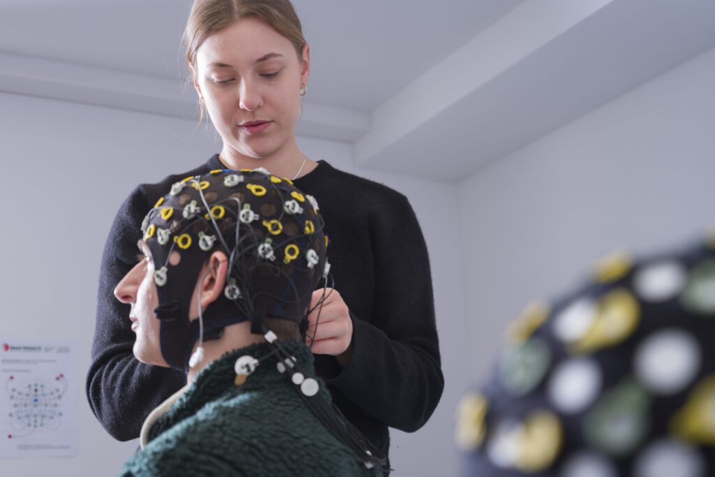
We are also interested in how memory encoding is shaped by different brain states. Here we are currently investigating different levels of information processing during transitional states between waking and sleeping with simultaneous EEG-fMRI. We are especially interested in the physiological factors that determine whether a certain component of information processing (e.g. stimulus perception, behavioral responsiveness, memory encoding) can be executed or not.
Imaging Human Neuroplasticity
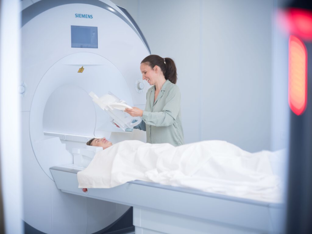
Functional MRI is a useful technique to image internal representations in the brain while they are activated. When it comes to memory, this implies that we can nicely assess mnemonic processing during encoding or recall. However, to infer the location in which mnemonic content is stored during offline phases, optimally we would like to assess learning-induced structural changes that indicate the generation or reorganization of neuronal connections.
Our lab is exploring possibilities to image human neuroplasticity noninvasively and in vivo with the help of MR-based biomarkers. In a first step, we could already show that the combination of functional and diffusion MRI can detect learning-induced microstructural plasticity rapidly after learning in task-relevant brain regions, which relates to subsequent memory performance.